
Rotator Cuff Weakness During Orthopedic Testing: What Are You Actually Feeling?
When a patient demonstrates weakness during resisted orthopedic testing, is that weakness caused by structural failure of the muscle-tendon unit, or is pain causing the nervous system to inhibit the muscle and reduce its ability to generate force?
The answer is often both, but the contribution of each can vary considerably.
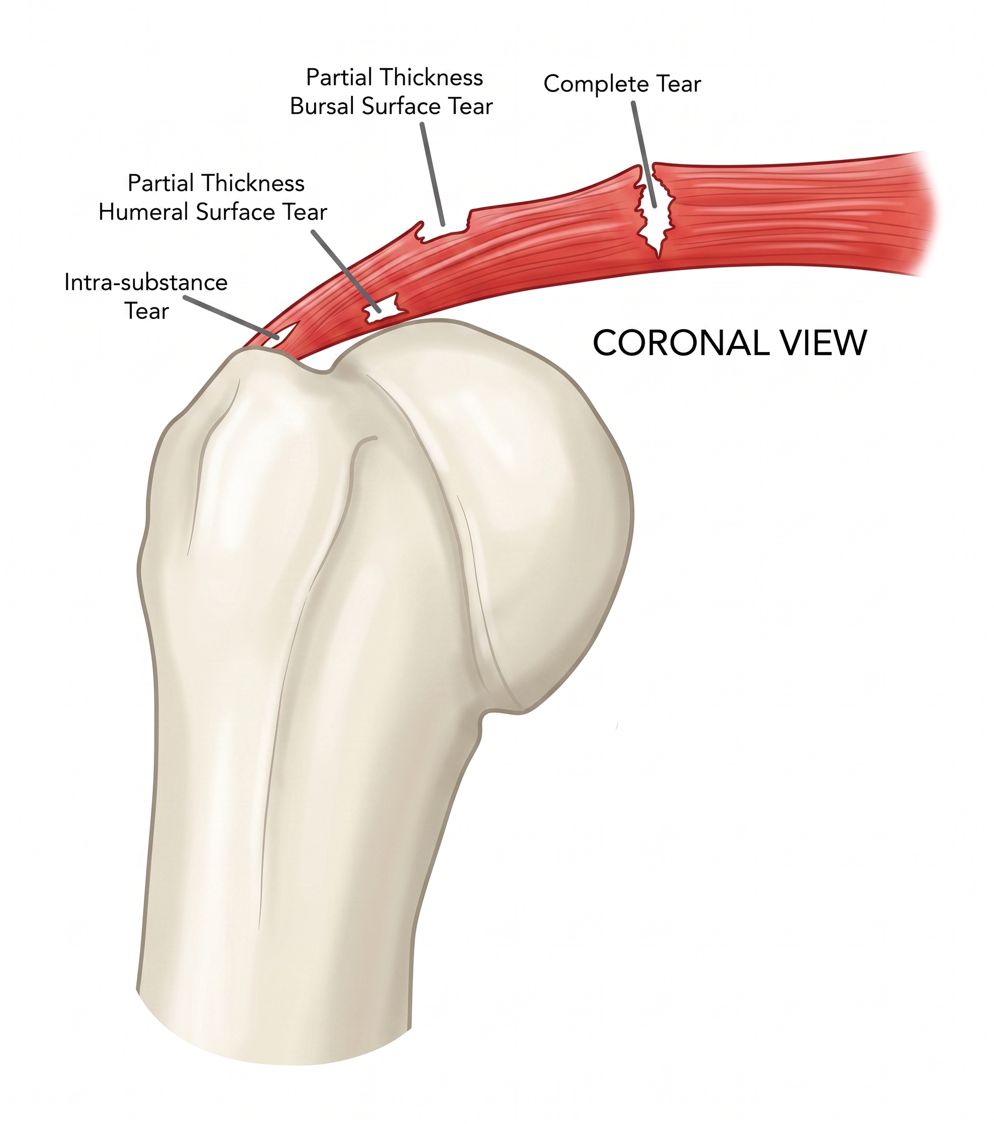
With tendinopathy or a partial-thickness tendon tear, the structural damage can reduce the tendon’s ability to transmit force efficiently. In other words, the muscle may be contracting appropriately, but some of that force is lost because the tendon is mechanically compromised.
Is The Weakness Neurological?
The weakness you perceive clinically may be greater than the structural damage alone would predict. Nociceptive input from the injured tissue can alter motor output through spinal and supraspinal mechanisms, producing protective inhibition and reducing voluntary motor-unit recruitment. The nervous system essentially places a temporary “governor” on force production.
This distinction is clinically important because neurophysiologic weakness can change rapidly, whereas structural weakness cannot.
But you already knew this… Your chiropractic treatments may produce immediate changes in pain, range of motion, and strength. So put it to the test. Perform your orthopedic strength testing before treatment, deliver your usual care, and then repeat the exact same test afterward. You may be surprised by how much more force the patient can immediately produce.
The tendon didn’t structurally repair itself in five minutes. So what changed?
Most likely, you altered some combination of pain, motor inhibition, biomechanics, confidence, and voluntary muscle recruitment. And that immediate change may tell you something important about the weakness you felt during your original examination.
Is The Weakness Structural?
With larger or chronic tendon tears, however, the mechanical component becomes increasingly important. Loss of tendon continuity, altered force transmission, muscle atrophy, fatty infiltration, and chronic changes in muscle architecture can create true mechanical weakness that cannot simply be “turned back on.”
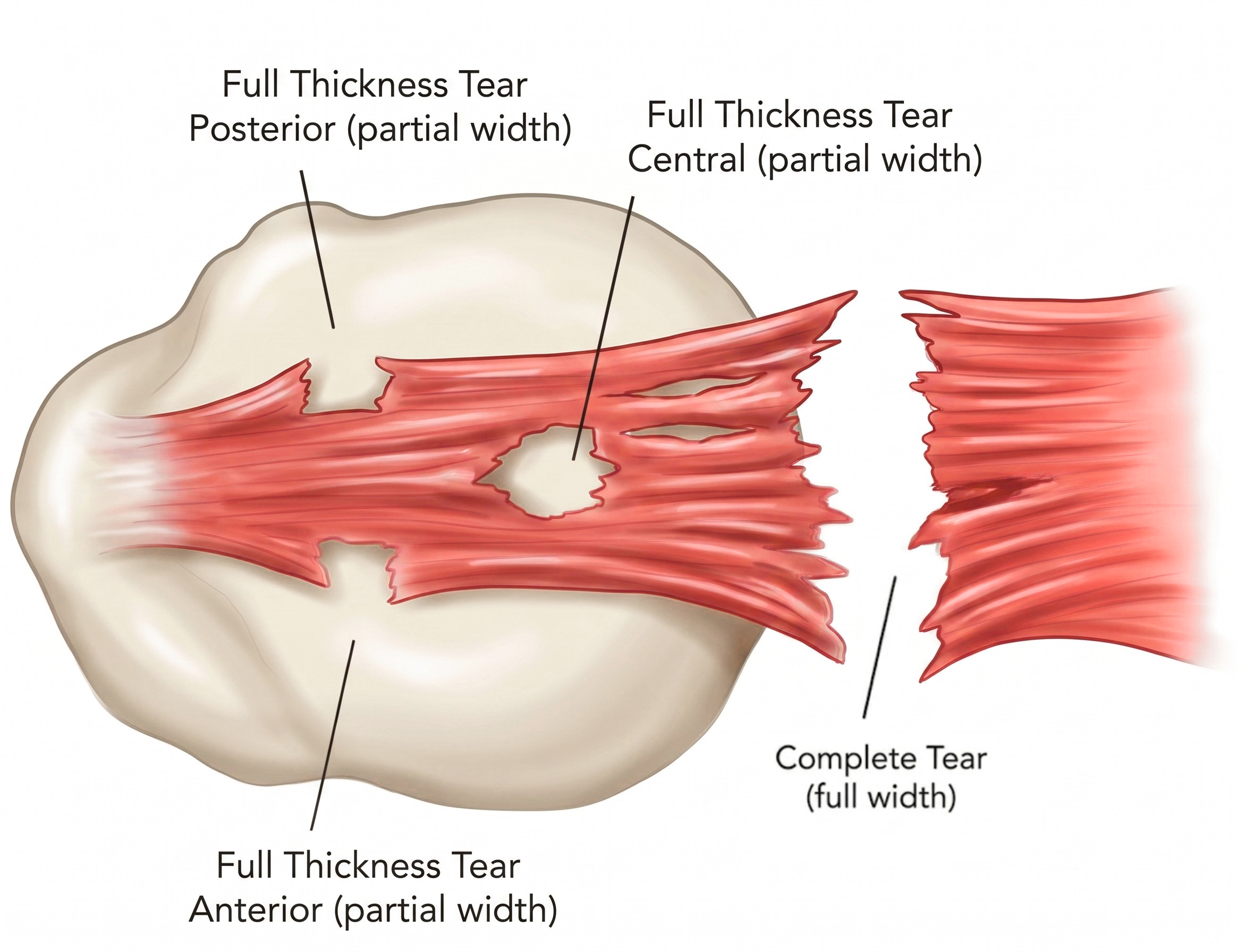
Clinical takeaway: When you feel weakness during an orthopedic test, don't automatically interpret it as a direct measurement of tissue damage. What you're testing is the output of the entire muscle-tendon-nervous-system complex. The weakness may represent a mixture of:
Structural compromise + pain-related inhibition + altered motor recruitment + biomechanics + chronic muscular changes
That is also why retesting after an intervention can be clinically useful. A rapid improvement in force suggests that at least part of the original weakness was modifiable rather than purely structural.
Putting This Into Practice: What Rotator Cuff Testing Can Teach Us About Weakness
When your patient presents with shoulder pain and weakness, can your orthopedic examination help you recognize what is actually happening within the muscle-tendon unit?
Is this rotator cuff tendinopathy with an intact tendon?
A partial-thickness tear producing pain and inhibition?
A chronic tear that has progressed to meaningful structural weakness?
Or a full-thickness tear that warrants imaging and potentially an orthopedic referral?
The difference matters because it alters our prognosis and treatment strategies. And ideally, we want to recognize that progression before the obvious lag sign appears. The challenge is understanding what the weakness beneath your hand is actually telling you.
For years, I have relied on three tests when evaluating patients with suspected full-thickness rotator cuff tears:
Dynamic Isotonic Manipulation Examination (DIME) for supraspinatus dysfunction
Internal Rotation Lag Sign for subscapularis dysfunction
External Rotation Lag Sign for infraspinatus and teres minor dysfunction
I still like these tests. In fact, I love these tests and use them for every single patient with a suspected shoulder injury. But I have changed how I think about when to use them.
These three tests are particularly useful when I am concerned about a significant rotator cuff tear, long-standing dysfunction, or weakness associated with more advanced tendon failure.
My Three Tests for Significant or Chronic Rotator Cuff Dysfunction
1. Dynamic Isotonic Manipulation Examination: Supraspinatus
The Dynamic Isotonic Manipulation Examination, or DIME, can be used to evaluate supraspinatus function and has demonstrated high sensitivity for full-thickness supraspinatus tears. I particularly like the ability to evaluate both pain and weakness.
Pain tells me one thing. True weakness tells me something different.
When a patient demonstrates substantial weakness rather than simply pain inhibition, my concern for more significant tendon involvement increases.

2. Internal Rotation Lag Sign: Subscapularis
The Internal Rotation Lag Sign is highly specific for significant subscapularis dysfunction, making a positive test clinically meaningful. However, its lower sensitivity means a negative test does not rule out a partial tear or earlier subscapularis pathology, as enough intact tendon may remain to maintain the position.
Keep in mind that the volume of the subscapularis is three times bigger than the other three rotator cuff muscles combined.
3. External Rotation Lag Sign: Infraspinatus and Teres Minor
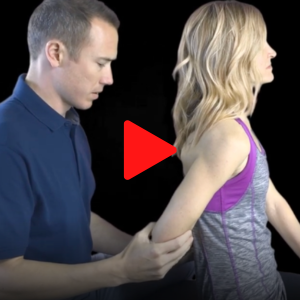
The External Rotation Lag Sign follows the same general principle for the posterior rotator cuff.
Can the patient maintain the externally rotated position? When they cannot, I become considerably more concerned about substantial infraspinatus or teres minor dysfunction. Again, this is a terrific test when positive, but it should not be expected to catch every early or partial-thickness tear.
Put Evidence-Based Care Into Practice
Want more evidence-based clinical insights like these? ChiroUp puts orthopedic testing, diagnosis, treatment protocols, exercises, and patient education resources at your fingertips, helping you turn the latest research into practical care.
Try it for free →There is a problem with testing only for weakness with a chiropractic exam. Think about the rotator cuff as a rope. A rope can be frayed without being broken. The same concept applies to a tendon. A patient may have tendinopathy or a partial-thickness tear while retaining sufficient functional tendon fibers to maintain relatively normal strength.
As the pathology progresses, however, the clinical picture can change. Pain may be followed by measurable weakness. With a larger or full-thickness tear, the patient may eventually lose the ability to maintain a specific shoulder position altogether. That is where lag signs become especially useful. A positive lag sign tells me something very different than pain during resisted testing.
What Do Chiropractors Need to Know About Partial-Thickness Rotator Cuff Tears?
This is where I believe chiropractors should consider changing their examination.
If my clinical question is: "Does this patient have early rotator cuff disease or a possible partial-thickness tear?"
It will require a more sensitive first layer of testing. My preferred three-test screen becomes:
1. Supraspinatus: Jobe / Empty Can Test
The Jobe test, commonly called the Empty Can test, provides a useful initial assessment of the supraspinatus. Rather than waiting for profound weakness or loss of function, I am looking for:
Pain
Weakness
Asymmetry
Reproduction of the patient's familiar symptoms
Research has demonstrated relatively high sensitivity for supraspinatus tears, making it useful as an initial screening maneuver. If Jobe testing is abnormal and DIME subsequently demonstrates substantial weakness, my level of concern changes.
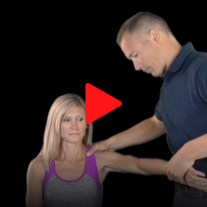
2. Subscapularis: Bear Hug Test
For earlier subscapularis involvement, I prefer the Bear Hug test over relying exclusively on an Internal Rotation Lag Sign. Research comparing common subscapularis tests has generally shown that these tests are highly specific but relatively insensitive. Among commonly used maneuvers, the Bear Hug has demonstrated better sensitivity than the Internal Rotation Lag Sign.
That makes it a better screening test. If the Bear Hug is abnormal, I can then perform the Internal Rotation Lag Sign to determine whether there is more substantial functional loss.

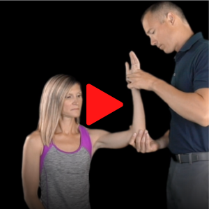
3. Infraspinatus/Teres Minor: Resisted External Rotation
For the posterior cuff, I begin with resisted external rotation. I am looking for pain, weakness, and side-to-side differences. If resisted external rotation demonstrates meaningful weakness, I can progress to the External Rotation Lag Sign.
If I suspect substantial teres minor involvement or a larger posterosuperior cuff tear, the Hornblower's Sign can provide additional information.
Clinical Pearl: The biggest change I’ve made in my approach to the rotator cuff examination is to think of it in two levels. Instead of asking, “What are the three best rotator cuff tests?” I now start with a more important question: “What clinical question am I trying to answer?”
Level 1: Is the Rotator Cuff Abnormal?
For earlier rotator cuff disease and possible partial-thickness involvement, consider:
Supraspinatus: Jobe / Empty Can
Subscapularis: Bear Hug
Infraspinatus/Teres Minor: Resisted External Rotation
These tests provide a broader screen for pain, weakness, and early dysfunction.
Level 2: Has a significant functional failure occurred?
When substantial weakness, chronicity, or a high-grade/full-thickness tear is suspected, consider:
Supraspinatus: DIME
Subscapularis: Internal Rotation Lag Sign
Infraspinatus/Teres Minor: External Rotation Lag Sign
Now you are asking a different question. The first group casts the net. The second group indicates whether something larger may be caught in it.
A Simple Rotator Cuff Testing Strategy for Chiropractors
| Rotator Cuff | Screen for Earlier Disease | Assess Significant Dysfunction |
|---|---|---|
| Supraspinatus | Jobe / Empty Can | DIME |
| Subscapularis | Bear Hug | Internal Rotation Lag Sign |
| Infraspinatus / Teres Minor | Resisted External Rotation | External Rotation Lag Sign |
Seven Surprising Facts You Probably Didn’t Know About Partial-Thickness Tendon Tears
-
The muscle fibers proximal to the tendon may initially retain essentially normal contractile machinery. Actin-myosin cross-bridge formation, motor-unit recruitment, and the muscle's theoretical capacity to generate tension can remain intact.
The problem is downstream.
The tendon is the series elastic force-transmission structure between muscle and bone. If some portion of its collagen-fascicle architecture is disrupted, the muscle can generate force, but the damaged tendon must transmit that force through a smaller and mechanically altered load-bearing structure.
-
This is the most important concept.
Imagine that a tendon contains many parallel, interconnected collagen fascicles that transmit load. If some are disrupted, they no longer participate normally in longitudinal load transmission. The remaining intact tissue, therefore, assumes a greater proportion of the applied load.
But a tendon is not a simple bundle of independent parallel cables, so you cannot say: "30% torn = 30% weaker."
That's physiologically incorrect.
There is interfascicular and matrix-mediated shear transfer, regional variation in material properties, nonlinear stress-strain behavior, differences in tear geometry, and complex load sharing between adjacent portions of a tendon and, in the rotator cuff, between neighboring cuff tendons. Models that simply distribute the lost load equally among the remaining fibers do not adequately capture the behavior of partially torn tendons. (1)
-
This is particularly relevant to the rotator cuff.
Experimental work on partial-thickness supraspinatus tears demonstrates that as tear depth increases, strain increases in the remaining intact tendon. In one cadaveric model, this became especially pronounced once the tear exceeded approximately 50% of tendon thickness. (2)
So conceptually:
Small tear → load redistribution may be well tolerated
Larger tear → less intact cross-sectional area + greater regional strain
Still larger tear → disproportionate strain on remaining fibers + reduced mechanical reserve
That helps explain why a person can have a genuine partial-thickness tear and still demonstrate surprisingly good strength during a clinical examination. Their tendon hasn't necessarily lost its ability to transmit the force demanded by your test. It has lost reserve capacity.
-
Suppose an intact supraspinatus tendon has substantially more load-bearing capacity than is required for a routine manual muscle test. A 20% partial tear might reduce its ultimate load capacity and stiffness, yet the remaining tendon could still transmit all of the force required during your clinical test. The patient, therefore, tests 5/5.
That does not mean the tendon is normal. It means:
The required load < remaining functional capacity.
As the demand rises, the deficit may become apparent. Heavy resistance, repetitive loading, fatigue, certain joint positions, or higher loading rates can expose a deficit that isn't apparent during a low-load examination.
-
So, measured weakness in a painful patient can contain at least two components:
Mechanical weakness from impaired force transmission. Tendinopathy without a discrete tear can alter the mechanical behavior of the tendon through changes in collagen organization, extracellular matrix, water content, cross-sectional morphology, and regional stiffness.
Pain-associated/neuromotor weakness from altered motor output. If loading produces pain, motor output may be modified through protective strategies, altered recruitment, reduced voluntary activation, or changes in force control. A recent systematic review found that musculoskeletal pain is, on average, associated with impaired force steadiness, although the magnitude and mechanisms vary substantially across conditions and individuals. (3)
-
Absolutely not. This is one of the most important clinical lessons in rotator cuff pathology. Structural pathology and pain correlate surprisingly poorly.
Population studies repeatedly demonstrate substantial numbers of completely asymptomatic rotator cuff tears, including full-thickness tears. One population study found that about 65% of identified full-thickness tears were asymptomatic; another, more recent cohort found nearly half were asymptomatic. (4)
Even more striking, asymptomatic individuals with complete supraspinatus tears can demonstrate measurable strength reductions while reporting no pain or functional limitation. (5)
So these are three separate variables: Structural integrity ≠ pain ≠ strength. They interact, but none is a perfect proxy for another.
-
Pain depends on considerably more than the percentage of collagen disrupted.
Potential contributors include local nociceptive signaling, inflammatory mediators, adjacent bursal involvement, mechanical compression, tendon loading characteristics, tear progression, sensitization, and individual nervous-system processing.
Tear size appears to have some relationship with symptoms at a population level, but it is far from deterministic. In one cohort, symptom severity was not related to structural severity until tears exceeded about 2.5 cm. (6)
Prospective research is also interesting here: when previously asymptomatic cuff tears become painful, this has been associated with tear enlargement and altered shoulder function, but the transition to pain did not necessarily produce measurable changes in external-rotation strength. (7)
That's a beautiful demonstration of why pain, structure, and strength cannot be treated as interchangeable measurements.
Summary: What Does Weakness Tell You About a Rotator Cuff Tear?
After digging into the research, I’ve changed how I interpret weakness during a rotator cuff exam.
Weakness is not the diagnosis; it is information.
A partial-thickness tear reduces the tendon’s load-bearing capacity, but structural damage does not predictably correlate with weakness or pain because the strength we feel reflects the entire muscle-tendon-neuromuscular system, including tendon integrity, cuff compensation, scapular mechanics, pain, and neuromuscular inhibition. This is why
I now approach testing in two levels: use tests such as Jobe, Bear Hug, and resisted external rotation to identify probable cuff involvement, then look for more substantial deficits, including lag-type findings, that increase suspicion for significant structural compromise and the need for imaging or referral.
And don’t forget to retest: if strength improves immediately after treatment, the tendon didn’t repair itself in five minutes; you likely changed pain, biomechanics, inhibition, or motor recruitment.
The real question isn’t simply, “Is this patient weak?” It’s, “What is this weakness telling me?”
ChiroUp helps you answer questions like these with evidence-based clinical resources at your fingertips. From orthopedic testing and diagnosis to treatment protocols, exercises, and patient education, ChiroUp helps you turn the latest research into practical care for the patients in front of you.
-
Pensalfini M, Duenwald-Kuehl S, Kondratko-Mittnacht J, Lakes R, Vanderby R. Evaluation of global load sharing and shear-lag models to describe mechanical behavior in partially lacerated tendons. J Biomech Eng. 2014 Sep;136(9):091006. doi: 10.1115/1.4027714. PMID: 24845861; PMCID: PMC4112931.
Yang S, Park HS, Flores S, Levin SD, Makhsous M, Lin F, Koh J, Nuber G, Zhang LQ. Biomechanical analysis of bursal-sided partial thickness rotator cuff tears. J Shoulder Elbow Surg. 2009 May-Jun;18(3):379-85. doi: 10.1016/j.jse.2008.12.011. Epub 2009 Mar 9. PMID: 19269860.
Arvanitidis M, Falla D, Sanderson A, Martinez-Valdes E. Does pain influence control of muscle force? A systematic review and meta-analysis. Eur J Pain. 2025 Feb;29(2):e4716. doi: 10.1002/ejp.4716. Epub 2024 Aug 23. PMID: 39176440; PMCID: PMC11671343.
Minagawa H, Yamamoto N, Abe H, Fukuda M, Seki N, Kikuchi K, Kijima H, Itoi E. Prevalence of symptomatic and asymptomatic rotator cuff tears in the general population: From mass-screening in one village. J Orthop. 2013 Feb 26;10(1):8-12. doi: 10.1016/j.jor.2013.01.008. PMID: 24403741; PMCID: PMC3768248.
Schibany N, Zehetgruber H, Kainberger F, Wurnig C, Ba-Ssalamah A, Herneth AM, Lang T, Gruber D, Breitenseher MJ. Rotator cuff tears in asymptomatic individuals: a clinical and ultrasonographic screening study. Eur J Radiol. 2004 Sep;51(3):263-8. doi: 10.1016/S0720-048X(03)00159-1. PMID: 15294335.
Hinsley H, Ganderton C, Arden NK, Carr AJ. Prevalence of rotator cuff tendon tears and symptoms in a Chingford general population cohort, and the resultant impact on UK health services: a cross-sectional observational study. BMJ Open. 2022 Sep 13;12(9):e059175. doi: 10.1136/bmjopen-2021-059175. PMID: 36100305; PMCID: PMC9472112.
Mall NA, Kim HM, Keener JD, Steger-May K, Teefey SA, Middleton WD, Stobbs G, Yamaguchi K. Symptomatic progression of asymptomatic rotator cuff tears: a prospective study of clinical and sonographic variables. J Bone Joint Surg Am. 2010 Nov 17;92(16):2623-33. doi: 10.2106/JBJS.I.00506. PMID: 21084574; PMCID: PMC2970889.