
Demystifying Modic Changes - Your Ultimate 5-minute Guide

This blog will answer a provider’s top 10 questions about Modic changes. In less than 5 minutes, you’ll better understand Modic classifications, causes, treatment options, and the truth about the relationship between Modic changes and pain. This straightforward evidence-based summary includes a tutorial video and two downloadable infographics.
1. What Are Modic Changes?
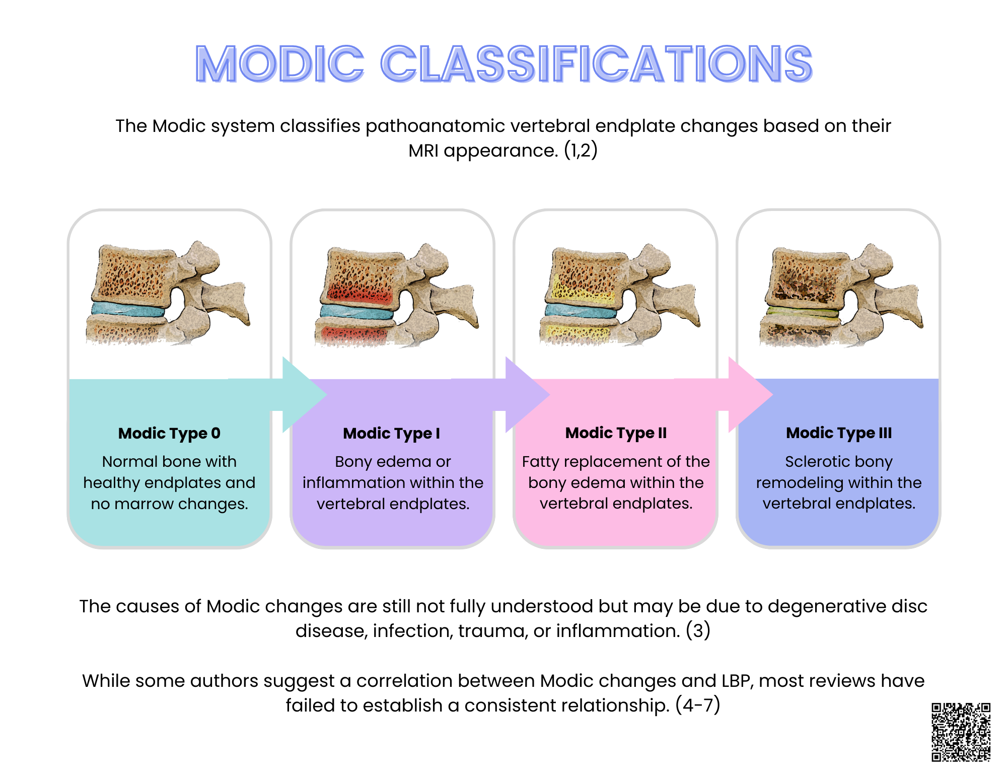
In 1988, Michael Modic, a neuroradiologist from the Cleveland Clinic, classified vertebral endplate changes into three types based on their T1 and T2 MRI appearances. (1-3)
Modic Type 1 MRI changes: T1 Low signal; T2 High signal
Modic Type 2 MRI changes: T1 high signal; T2 Iso to High signal
Modic Type 3 MRI changes: T1 Low signal; T2 Low signal

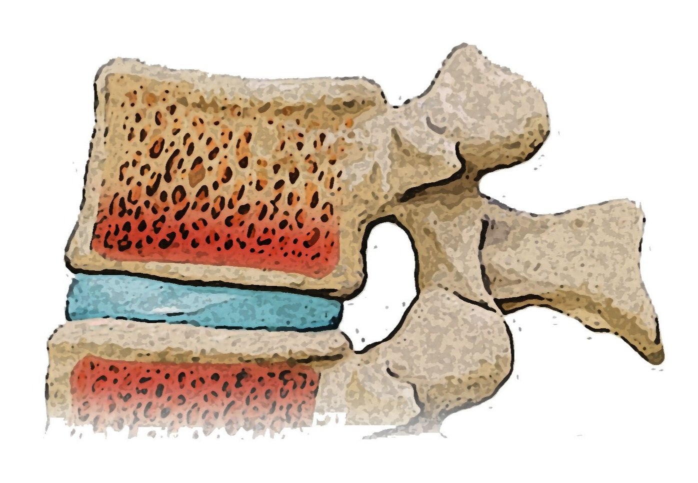
2. What Are Modic Type 1 Endplate Changes?
Type 1 Modic changes include bony edema or inflammation within the vertebral endplates.
3. What Causes Modic Type 1 Changes?
The underlying causes of Modic Changes type 1 are not fully understood but are theorized to be triggered by some combination of injury, inflammation, or infection. (4,5)
Trauma- Mechanical Modic changes might occur due to trauma causing micro-fractures within the endplate and vertebral bone. This can lead to bleeding, edema, and the development of vascular granulation tissue adjacent to the endplates. (2,3)
Inflammation- An alternative explanation for developing Modic changes is that the nuclear tissue invades the highly vascular marrow through endplate microfractures. Because this nuclear material has not previously been exposed to the blood and immune systems, an inflammatory autoimmune response ensues. (6)
Infection- Another theorized cause of Modic changes is due to a bacterial infection in the adjacent intervertebral disc. (7-12) Multiple studies have demonstrated the presence of ongoing bacterial infection in approximately 30-34% of disc lesion patients. (21,22)
Check out this short tutorial on the causes of Modic change.
4. What Are Modic Type 2 Endplate Changes?
Type 2 Modic changes signify the replacement of the Modic Type 1 bony edema with fat.

5. What Causes Modic Type 2 Changes?
Modic changes type 2 represent the conversion of normal red bone marrow into yellow fatty marrow, likely due to marrow ischemia. (13)
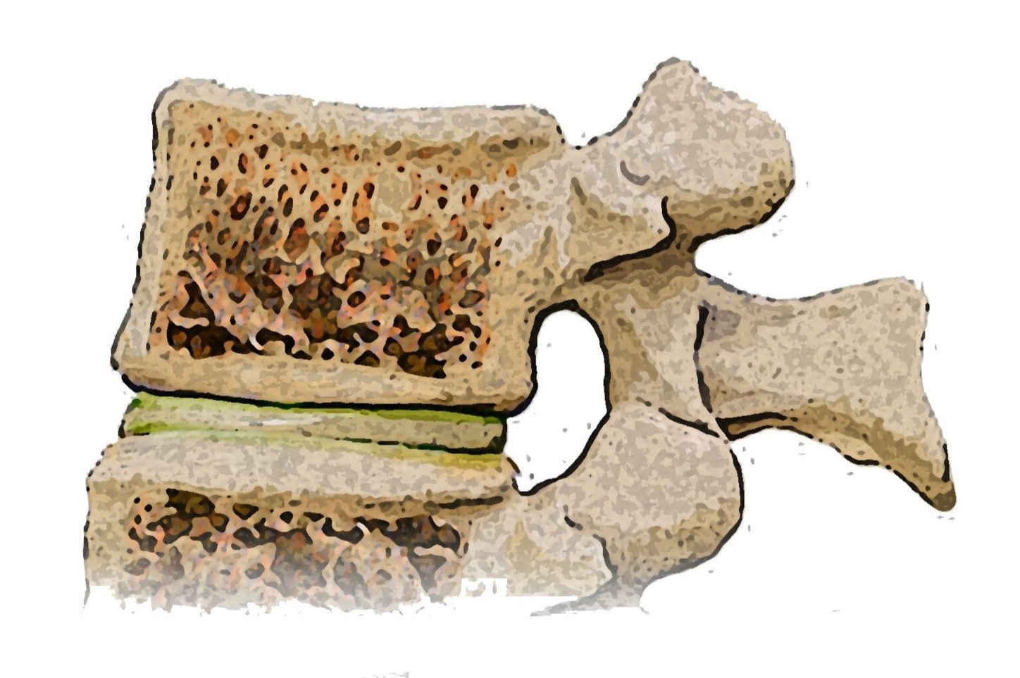
6. What Are Modic Type 3 Endplate Changes?
Type 3 Modic changes involve sclerotic bony remodeling within the vertebral endplates.
7. What Causes Modic Type 3 Changes?
Modic changes type 3 refers to the hardening or thickening of the bone beneath the cartilage of the vertebral endplate. Like other Modic changes, the exact cause of Modic type 3 change is not fully known but is likely a result of the body's attempt to repair and stabilize the affected segments.
Subscribers can download ChiroUp’s newest infographic, detailing Modic Changes in the forms library.
Transform your practice with ChiroUp - Try it risk-free!
8. What Is The Relationship Between Modic Changes And Pain?
Patients will sometimes ask, “Are Modic changes painful?” The short answer is “not necessarily.” AKA, there’s a longer answer :-)
Inflammatory bony changes, specifically of Modic type 1, possess the inherent potential to trigger pain. And some early papers confirmed a link between Modic endplate changes and back pain, particularly nighttime pain and morning stiffness (14,15). However, several more recent reviews indicate no association between Modic changes and LBP:
“Modic changes in the lumbar spine are not associated with clinically significant axial low back pain severity or patient disability.” (16)
“The associations between Modic changes and LBP-related outcomes are inconsistent.” (17)
“There is no conclusive evidence on the causative role of Modic change in chronic low back pain or any influence on the long-term outcome in patients with LBP or lumbar disc herniations.” (18)
“The inconsistency of findings … means that no conclusions can be drawn about an association between Modic change and LBP.” (19)
9. How Are Modic Changes Treated?
Any MRI finding is potentially concerning to patients, evidenced by the fact that two common internet searches are:
What is the modic type 1 endplate changes treatment?
What is the modic type 2 endplate changes treatment?
Modic changes, like joint degeneration and disc lesions, are pathoanatomic indicators of persistent physical stress on the spine. While these structural abnormalities may not directly cause symptoms, the underlying stressors contributing to Modic changes' development can also lead to concurrent complaints. Effective resolution of symptoms involves addressing and eliminating the underlying sources of stress on the spine. By targeting and removing these stressors, it is possible to alleviate the associated symptoms, despite the structural changes.
And just in case you’re a patient who stumbled upon this top chiropractic blog, you should know two things:
Having Modic changes or degeneration (arthritis) does not necessarily mean you’re doomed to a lifetime of pain.
Conservative chiropractic care is a potent, medically-recommended option for back pain sufferers, even those with degeneration or arthritis. (20)
Want to learn more? Check out this Natural Solutions blog, detailing a safe, effective, drug-free, non-surgical option for back pain and other complaints.
10. How Do I Educate Patients About Endplate Modic Changes?
ChiroUp has you covered! Subscribers can download the lay infographic for Modic Changes from their forms library 24/7. (Visit Practice Resources> Forms library> and Search “Modic”)

-
1. Modic MT, Masaryk TJ, Ross JS, Carter JR. Imaging of degenerative disk disease. Radiology. 1988 Jul;168(1):177-86. Link
2. Modic MT, Steinberg PM, Ross JS, Masaryk TJ, Carter JR. Degenerative disk disease: assessment of changes in vertebral body marrow with MR imaging. Radiology. 1988 Jan;166(1):193-9. Link
3. Gaillard F, Hapugoda S, Murphy A, et al. Modic type I endplate change. Reference article, Radiopaedia.org (Accessed on 14 Jun 2023) from Link
4. Din RU, Cheng X, Yang H. Diagnostic role of magnetic resonance imaging in low Back pain caused by vertebral endplate degeneration. Journal of Magnetic Resonance Imaging. 2022 Mar;55(3):755-71. Link
5. Crockett MT, Kelly BS, van Baarsel S, Kavanagh EC. Modic type 1 vertebral endplate changes: injury, inflammation, or infection?. American Journal of Roentgenology. 2017 Jul;209(1):167-70. Link
6. Crock HV. The presidential address: ISSLS: Internal disc disruption a challenge to disc prolapse fifty years on. Spine. 1986 Jul 1;11(6):650-3. Link
7. Hirabayashi S, Kumano K, Tsuiki T, Eguchi M, Ikeda S. A dorsally displaced free fragment of lumbar disc herniation and its interesting histologic findings: a case report. Spine. 1990 Nov;15(11):1231-3. Link
8. Ito T, Yamada M, Ikuta F, Fukuda T, Hoshi SI, Kawaji Y, Uchiyama S, Homma T, Takahashi HE. Histologic evidence of absorption of sequestration-type herniated disc. Spine. 1996 Jan 15;21(2):230-4. Link
9. Doita M, Kanatani T, Harada T, Mizuno K. Immunohistologic study of the ruptured intervertebral disc of the lumbar spine. Spine. 1996 Jan 15;21(2):235-41. Link.
10. Grönblad M, Virri J, Tolonen J, Seitsalo S, Kääpä E, Kankare J, Myllynen P, Karaharju EO. A controlled immunohistochemical study of inflammatory cells in disc herniation tissue. Spine. 1994 Dec 15;19(24):2744-51. Link
11. Urquhart DM, Zheng Y, Cheng AC, Rosenfeld JV, Chan P, Liew S, Hussain SM, Cicuttini FM. Could low grade bacterial infection contribute to low back pain? A systematic review. Bmc Medicine. 2015 Dec;13:1-3. Link
12. Ganko R, Rao PJ, Phan K, Mobbs RJ. Can bacterial infection by low virulent organisms be a plausible cause for symptomatic disc degeneration? A systematic review. Spine. 2015 May 15;40(10):E587-92. Link
13. Radswiki T, Hacking C, Yap J, et al. Modic type endplate changes. Reference article, Radiopaedia.org (Accessed on 14 Jun 2023) from Link
14. Bailly F, Maigne JY, Genevay S, Marty M, Gandjbakhch F, Rozenberg S, Foltz V. Inflammatory pain pattern and pain with lumbar extension associated with Modic 1 changes on MRI: a prospective case–control study of 120 patients. European spine journal. 2014 Mar;23:493-7. Link
15. Arnbak B, Jurik AG, Jensen TS, Manniche C. Association between inflammatory back pain characteristics and magnetic resonance imaging findings in the spine and sacroiliac joints. Arthritis care & research. 2018 Feb;70(2):244-51. Link
16. Lambrechts MJ, Issa TZ, Toci GR, Schilken M, Canseco JA, Hilibrand AS, Schroeder GD, Vaccaro AR, Kepler CK. Modic Changes of the Cervical and Lumbar Spine and Their Effect on Neck and Back Pain: A Systematic Review and Meta-Analysis. Global Spine Journal. 2022 Nov 30:21925682221143332. Link
17. Herlin C, Kjaer P, Espeland A, Skouen JS, Leboeuf-Yde C, Karppinen J, Niinimäki J, Sørensen JS, Storheim K, Jensen TS. Modic changes—their associations with low back pain and activity limitation: a systematic literature review and meta-analysis. PloS one. 2018 Aug 1;13(8):e0200677. Link
18. Viswanathan VK, Shetty AP, Rajasekaran S. Modic changes-An evidence-based, narrative review on its patho-physiology, clinical significance and role in chronic low back pain. Journal of Clinical Orthopaedics and Trauma. 2020 Sep 1;11(5):761-9. Link
19. Hopayian K, Raslan E, Soliman S. The association of modic changes and chronic low back pain: A systematic review. Journal of Orthopaedics. 2022 Nov 17. Link
20. Qaseem A, Wilt TJ, McLean RM, Forciea MA, Clinical Guidelines Committee of the American College of Physicians. Noninvasive treatments for acute, subacute, and chronic low back pain: a clinical practice guideline from the American College of Physicians. Annals of internal medicine. 2017 Apr 4;166(7):514-30. Link
21. Capoor MN, Ruzicka F, Schmitz JE, James GA, Machackova T, Jancalek R, Smrcka M, Lipina R, Ahmed FS, Alamin TF, Anand N. Propionibacterium acnes biofilm is present in intervertebral discs of patients undergoing microdiscectomy. PLoS One. 2017 Apr 3;12(4):e0174518. Link
22. Ohrt‐Nissen S, Fritz BG, Walbom J, Kragh KN, Bjarnsholt T, Dahl B, Manniche C. Bacterial biofilms: a possible mechanism for chronic infection in patients with lumbar disc herniation–a prospective proof‐of‐concept study using fluorescence in situ hybridization. Apmis. 2018 May;126(5):440-7. Link