Clinical Findings: The Top 6 Threatening Spinal Diagnoses
Repeatedly hearing familiar presentations for common diagnoses can lull clinicians into a false sense of security. Even astute clinicians develop a bias toward statically probable benign neuromusculoskeletal diagnoses. Unfortunately, this complacency can lead to disastrous outcomes when a more threatening problem masquerades as something less serious.
A recent American Journal of Medicine systematic review of 22 studies involving 41,320 patients concluded that up to 5% of LBP patients presenting to an emergency department have a serious underlying pathology (fracture, infection, cancer, cauda equina, etc.) (1) Prior studies have suggested that the same threatening diagnosis present to primary care offices at a rate of approximately 1%. (2)
Spinal compressive disorders rank high on the list of back pain emergencies due to the threat of cord ischemia and nerve cell death. This blog will cover the six most common spinal-compressive diagnoses and the clinical findings that can alert us to these ominous problems- including a video tutorial with at tip so that you’ll never forget what UMNL pathology looks like.
“Atraumatic spinal emergencies often present a diagnostic and management dilemma for healthcare practitioners. Spinal epidural abscess, cauda equina syndrome, and spinal epidural hematoma are conditions that can insidiously present to outpatient medical offices. Unless a high level of clinical suspicion is maintained, these clinical entities may be initially misdiagnosed and mismanaged. Permanent neurologic sequela and even death can result if delays in appropriate treatment occur.”(4)
A 2019 focused literature review of 34 peer-reviewed studies highlighted four threatening spinal-compressive clinical scenarios that require emergent attention.
* Unless otherwise noted, all bullet points in items 1- 4 are from Babu et al.: (4)
1. Cervical myelopathy
Compression of the spinal cord within the central canal.
Common presenting neurologic signs and symptoms:
bilateral paresthesias, numbness, and altered proprioception
bilateral weakness, spasticity, or increased muscle tone
possible altered hand dexterity, gait, and balance difficulty
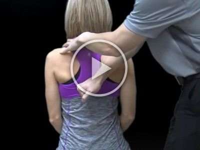
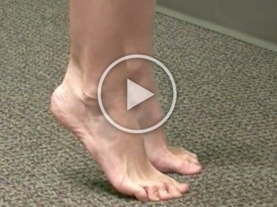
Upper motor neuron signs (e.g. hyperreflexia, Hoffman’s sign, inverted radial reflex, clonus, or Babinski) correlate with cervical cord dysfunction but are not always present in the setting of acute cord compression. (4)
Watch Dr. Bertelsman demonstrate how to evaluate for signs of cord compression, including a quick way to always remember which limb movements signify an upper motor neuron lesion.
2. Cauda equina syndrome
Compression of the neural elements caudal to the distal extent of the spinal cord.
The two most common presenting symptoms:
severe lower back pain (83-100% of cases)
unilateral or bilateral sciatica (90-100% of cases).
Additional ‘red-flags’ include:
motor or sensory deficits in one or both legs
saddle anesthesia
bowel, bladder, or sexual dysfunction
Clinical suspicion of cauda equina syndrome should prompt emergent MRI and spine surgical consultation. (4)
3. Spinal epidural abscess
Infection withaccumulation of pus in the epidural space that can compress the cord or nerve roots.
May present with radiculopathy or myelopathy depending on the location of the abscess.
Axial spine pain (72%) and fever (55%) are the most common symptoms. (3)
However, the full ‘classic triad’ for spinal epidural abscess consisting of fever, axial pain, and neurologic deficit is only seen in 8% of patients.
Common presenting comorbidities for spinal epidural abscess include (3,4):
previous focus of infection (22-54%)
diabetes (22-51%)
IV drug abuse (29-39%)
renal disease (7-37%)
Many patients who develop epidural abscesses have no risk factors
Lab shows elevated ESR & CRP, possible leukocytosis
Evaluate with full spine contrast MRI or myelogram (4)
4. Spinal epidural hematoma
Bleeding into the epidural space with subsequent compression of the neural elements.
Axial spine pain is the most common initial presenting symptom.
Patients may also complain of variable:
radicular pain
paresis or paralysis
myelopathy and bowel or bladder dysfunction
Most common in the cervicothoracic region
Suspicion should be high anytime a patient has severe spinal pain and a history of trauma, invasive spinal procedure, peri-spinal injections, epidural catheter, coagulopathy, or ongoing use of blood-thinning medications.
MRI is the gold standard for evaluation of spinal epidural hematoma. (4)
Here are two more common diagnoses, not detailed in the AJM paper, that also pose a compressive threat to the cord and nerve roots:
5. Compression fracture
vertebral body collapse posing a threat to the spinal cord and nerve roots.
History of variable trauma
Limited spinal range of motion (5)
Localized tenderness over the site of acute compression (7)
The inability to lie supine on an exam table with only one pillow supporting the head (Supine Sign) has relatively high sensitivity (81%) and specificity (93%) for vertebral compression fracture. (6)
Seated Closed Fist Percussion of the spine has a sensitivity and specificity near 90% for vertebral compression fracture. (6)
The Heel Drop Test may increase pain originating from a compression fracture. (8)
The most common sites for osteoporotic compression fracture are T6-T8, T11-L1, and L4. (9,10)
6. Neoplasm
new and abnormal tissue growth with compressive potential.
Metastatic lesions are responsible for about 85% of neoplastic spinal cord compression cases (12)
Metastatic seeding involves:
thoracic spine (70% of cases)
lumbar spine (20% of cases)
cervical spine (10% of cases)
Multiple spinal levels are affected in about 30% of patients. (12,13)
A high index of suspicion may be required when evaluating a patient with back pain and a history of malignancy, particularly: breast, prostate, kidney, lung, lymphoma, sarcoma, or multiple myeloma. (1)
Clinical signs might include spinal pain and tenderness with sensory loss, motor weakness, hyperreflexia (below lesion), and ataxia.
Signs of space-occupying lesion could include: Valsalva, Soto-hall, Slump test
Bottom Line: “If it doesn’t look, smell, or feel right- go with your gut and investigate further. As a clinician, it’s usually better to over-respond than under-respond. Looking for the worst possible scenario and being surprised by a benign diagnosis is always more comfortable than closing a relationship the other way around.”
While these six license-stealing threats may be top-of-mind at the moment, our recall will undoubtedly fade over time and in direct proportion to our preoccupation with “routine” care. That’s why it’s essential to have systems that increase our odds of identifying threats whenever they present. Check out this updated ChiroUp health history form that helps you collect essential information for identifying threats and directing care. Then, make sure you have a system ensuring that someone is responsible for collecting and reviewing this data for every new presentation.
It is crucial to stay up-to-date and equipped with essential knowledge. This year, I urge you to commit to continually refining your clinical skills—and with ChiroUp, it really doesn’t get any easier to stay on your A-game.
ChiroUp is a community committed to delivering the best possible care to every patient, every time—backed by evidence. We want you to join our growing alliance!
-
Galliker G, Scherer DE, Trippolini MA, Rasmussen-Barr E, LoMartire R, Wertli MM. Low Back Pain in the Emergency Department: Prevalence of Serious Spinal Pathologies and Diagnostic Accuracy of Red Flags–A Systematic Review. The American journal of medicine. 2019 Jul 3.
Henschke N, Maher CG, Refshauge KM, Herbert RD, Cumming RG, Bleasel J, York J, Das A, McAuley JH. Prevalence of and screening for serious spinal pathology in patients presenting to primary care settings with acute low back pain. Arthritis & Rheumatism: Official Journal of the American College of Rheumatology. 2009 Oct;60(10):3072-80.
Yusuf M, Finucane L, Selfe J. Red flags for the early detection of spinal infection in back pain patients. BMC Musculoskeletal Disorders. 2019 Dec;20(1):1-0.
Babu JM, Patel SA, Palumbo MA, Daniels AH. Spinal emergencies in primary care practice. The American journal of medicine. 2019 Mar 1;132(3):300-6.
Papa JA. Conservative management of a lumbar compression fracture in an osteoporotic patient: a case report. J Can Chiropr Assoc. 2012 Mar; 56(1): 29–39.
Langdon J, Way A, Heaton S, Bernard J, Molloy S. Vertebral compression fractures – new clinical signs to aid diagnosis. Annals of The Royal College of Surgeons of England. 2010;92(2):163-166.
Lee YL, Yip KM. The osteoporotic spine. Clin Orthop 1996;323: 91–7.
McGill S. Low Back Disorders: Evidence-based Prevention and Rehabilitation. Human Kinetics, 2007. p. 192
Haczynski J, Jakimiuk A. Vertebral fractures: a hidden problem of osteoporosis. Med Sci Monit. 2001 Sep-Oct;7(5):1108–17.
Patel U, Skingle S, Campbell GA, Crisp AJ, Boyle IT. Clinical profile of acute vertebral compression fractures in osteoporosis. Br J Rheumatol. 1991;30:418–421.
Schellinger KA, Propp JM, Villano JL, McCarthy BJ. Descriptive epidemiology of primary spinal cord tumors. Journal of neuro-oncology. 2008 Apr 1;87(2):173-9.
Borke J et al. Spinal Cord Neoplasms Clinical Presentation. MedScape. Accessed on 1/12/20 from: https://emedicine.medscape.com/article/779872-overview
Spinazze S, Caraceni A, Schrijvers D. Epidural spinal cord compression. Crit Rev Oncol Hematol. 2005 Dec. 56(3):397-406.


