
Chiropractic Evaluation: Carpal Tunnel vs. Pronator Teres
A recent prospective cohort study of more than 1100 patients identified the most sensitive and specific findings to establish the diagnosis of carpal tunnel syndrome (CTS).
The study found that “paresthesia in a median nerve distribution with nocturnal awakening” was 77% sensitive for the diagnosis of CTS. So where do the other 23% of cases arise? Check out this week’s chiropractic clinical blog for a quick yet potent review of the work-up for this common presentation.
When a patient presents with numbness and tingling in their first three and a half fingers, most clinicians presume a diagnosis of carpal tunnel syndrome. Without confirming this source of median nerve compression, we may be directing our treatment at the wrong culprit. Check out the following video tutorial of the most sensitive and specific tests for differentiating CTS from its imitators, including the often-overlooked pronator teres syndrome.
1. Etiology
Pronator Teres Syndrome describes the constellation of signs and symptoms resulting from compression of the median nerve by the pronator teres muscle near the elbow. It is the second most frequent cause of median nerve compression with features similar to but discernible from its more common distal counterpart, carpal tunnel syndrome. (1) Pronator Syndrome (PS) responsible for 9.2% of all cases of median nerve entrapment. (2).

Pronator syndrome (PS) is associated with prolonged or repetitive forearm pronation and finger flexion, i.e., gripping with the palm down. Carpenters, mechanics, assembly line workers, tennis players, rowers, and weightlifters are predisposed to this problem. (6) The condition is often associated with excessively developed forearm muscles and is more common in the dominant arm. (7,8) The typical age of onset is in the fifth decade, and the condition is four times more common in women than in men. (9) Like other neuropathies, patients with diabetes, alcoholism, or hypothyroidism are predisposed to this disorder.
2. Presentation
Pronator syndrome often presents as an aching discomfort in the volar forearm with associated paresthesias into the first three-and-a-half digits. Symptoms of pronator syndrome mimic those of carpal tunnel syndrome with some important points of differentiation:
Nocturnal exacerbations are common in carpal tunnel syndrome but notably absent in pronator syndrome. (10, 11,12)
Both are exacerbated by wrist flexion, but pronator syndrome symptoms are often increased with resisted or repetitive forearm pronation or supination.
Most significantly, isolated carpal tunnel syndrome symptoms do not affect the palm, whereas pronator teres syndrome usually will. (See graphic below)

Sensation to the first 3 ½ digits is supplied by the median nerve (green). However, the palm is innervated by the palmar branch of the median nerve (blue).
The palmar branch splits off proximally and travels outside of the carpal tunnel, meaning it would not be affected by compression within the tunnel.
3. Assessment
Last year’s AAOS study (18) defined the sensitivity of two tests commonly used to diagnose carpal tunnel syndrome:
Phalen sign sensitivity was 52.8%
Tinel sign (at the wrist) sensitivity was 37.7%
The chiropractic clinical evaluation of isolated pronator teres syndrome patients differs in the following ways:
Tinel sign is typically absent at the wrist but may be positive over the proximal anterior forearm
Phalen’s test is usually negative
Palpation demonstrates tenderness over the pronator teres and likely over the medial epicondyle
Orthopedic testing can help differentiate the site of involvement by generating tension on specific potential sites of median nerve compression.
Pronator Compression Test
The pronator compression test is positive when symptoms are reproduced within 30 seconds of applying deep, sustained compression of the pronator muscle. The uninvolved arm should remain asymptomatic. A small study by Gainor demonstrated 100% sensitivity for this test. (13)
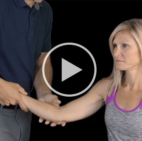
Pronator Teres Syndrome Test
The pronator teres syndrome test is performed with the patient’s elbow in 90 degrees of flexion. The practitioner stabilizes the elbow with one hand and asks the patient to pronate his hand against the practitioner’s resistance. The clinician extends the patient’s elbow while holding this resistance. Reproduction of symptoms suggests involvement of the pronator teres muscle.

Clinicians should be alert to the possibility of a “double crush syndrome,” wherein the median nerve is sensitized from axonal irritation at multiple sites. (14) Common double crush partners (and differential diagnosis considerations) for median nerve compression include cervical arthropathy, cervical disc, brachial plexus neuritis, TOS, and carpal tunnel syndrome.
4. Management
Fortunately, both CTS and PS respond well to conservative chiropractic management. A July 2018 systematic review comparing surgical vs. non-surgical (i.e., splint, steroid injection, or physical therapy) outcomes for CTS patients found: “No significant differences at 3 or 12 months” in terms of functional status, symptom severity, and nerve conduction outcomes. (17) And for pronator teres syndrome, another study demonstrated that 50% of patients reported resolution of symptoms within four months of initiating care.
An effective conservative management strategy for PS includes rest, median nerve flossing, manipulation, and myofascial release. Good clinical judgment is required to assess the point at which the benefits of soft tissue mobilization outweigh the risks of symptom exacerbation. When the symptoms are no longer acute, stretching and myofascial release of hypertonic pronator teres and wrist flexor muscles is appropriate. Stretching should be performed before nerve mobilization. Nerve mobilization should not proceed past the point of symptom provocation or exacerbation.
Perhaps the most crucial aspect of treatment is avoiding repetitive forceful gripping. A discussion of workstation ergonomics and specifically avoiding repetitive tasks is essential.
If every presentation of “paresthesia in a median nerve distribution with nocturnal awakening” results in a diagnosis of carpal tunnel syndrome, our outcome score is 77% – basically a low “C”.
Being able to successfully identify and manage non-CTS causes of hand paresthesia is essential in the new fee-for-outcomes world.
ChiroUp subscribers have 24/7 access to current best practice synopses for CTS, PS, and more than 100 other conditions. To access them, simply log in and navigate to Clinical Skills/ Condition References.
Not yet a subscriber? Start accessing ChiroUp resources today!
“This is probably THE best product I’ve ever taken a chance on. Its value can be recognized immediately. Great job guys!” – Dr. Williams | Creek Stone Integrated Care
-
Lee MJ, LaStayo PC. Pronator Syndrome and Other Nerve Compressions That Mimic Carpal Tunnel Syndrome. J Orthop Sport Phys Ther 2004;34:601-609.
Gessini L, Jandolo B, Pietrangeli A. Entrapment neuropathies of the median nerve at and above the elbow. Surg Neurol. 1983;19:112-116.
Eversmann WW. Proximal median nerve compression. Hand Clin. 1992;8:307-315.
Nebot-Cegarra J, Perez-Berruezo J, Reina de la Torre F. Variations of the pronator teres muscle: predispositional role to median nerve entrapment. Arch Anat Histol Embryol. 1991;74:35-45.
Tulwa N, Limb D, Brown RF. Median nerve compression within the humeral head of pronator teres. J Hand Surg [Br]. 1994;19:709-710.
Howard FM. Compression neuropathies in the anterior forearm. Hand Clin. 1986;2:737-745.
www.orthobullets.com/hand/6020/pronator-syndrome
www.mdguidelines.com/pronator-syndrome
Tetro AM, Pichora DR. High median nerve entrapments. An obscure cause of upper-extremity pain. Hand Clin. 1996;12:691-703.
Haussmann P, Patel MR. Intraepineurial constriction of nerve fascicles in pronator syndrome and anterior interosseous nerve syndrome. Orthop Clin North Am. 1996;27:339-344.
Rehak DC. Pronator syndrome. Clin Sports Med. 2001;20:531-540.
Werner CO, Rosen I, Thorngren KG. Clinical and neurophysiologic characteristics of the pronator syndrome. Clin Orthop. 1985;231-236.
Gainor BJ. The pronator compression test revisited. A forgotten physical sign. Orthop Rev. 1990;19:888-892.
Upton AR, McComas AJ. The double crush in nerve entrapment syndromes. Lancet 1973 Aug 18;2(7825):359-62.
Hartz CR, Linscheid RL, Gramse RR, Daube JR. The pronator teres syndrome: compressive neuropathy of the median nerve. J Bone Joint Surg Am. 1981 Jul;63(6):885-90.
Ebenbichler GR, Resch KL, Nicolakis P, et al. Ultrasound treatment for treating the carpal tunnel syndrome: randomised ‘‘sham’’ controlled trial. BMJ. 1998;316:731-735.
Qiyun S. et al. Comparison of the Short-term and Long-term Effects of Surgery and Nonsurgical Intervention in Treating Carpal Tunnel Syndrome: A Systematic Review and Meta-analysis. Hand. 2018 Jul 1:1558944718787892. [Epub ahead of print]
Hegmann KT et al. Median Nerve Symptoms, Signs, and Electrodiagnostic Abnormalities Among Working Adults. J Am Acad Orthop Surgery. 2018 Jul 19. [Epub ahead of print]